Leg Bones Diagram Labeled : Pelvis Definition Anatomy Diagram Facts Britannica : Click and start learning now!
Get link
Facebook
X
Pinterest
Email
Other Apps
Leg Bones Diagram Labeled : Pelvis Definition Anatomy Diagram Facts Britannica : Click and start learning now!. Your leg bones are the longest and strongest bones in your body. The leg consists of two long bones, the tibia and fibula, and the sesamoid bone, the patella, that serves as the knee cap. Bones are very busy even when you are sleeping at night. To understand one of the most complex joints of our body i.e. The knee joint is the largest joint in the body and is primarily a hinge joint, although some sliding and rotation occur.
There also are bands of fibrous connective tissue—the ligaments and the tendons—in intimate relationship with the parts of the a diagram of the human skeleton showing bone and cartilage. Virtual bone labwe need our bones to walk, run, jump and move, but this is not all they do. (a) tarsus of a dog in dorsal.,schematic drawing of the tarsal joint with the locations of different these pictures of this page are about:tarsal bones diagram. The leg consists of two long bones, the tibia and fibula, and the sesamoid bone, the patella, that serves as the knee cap. The bones mentioned in each human skeleton chart are:
Femur Definition Function Diagram Facts Britannica from cdn.britannica.com Learn more about the leg and knee anatomy by taking our special quiz, customized to focus on bones, muscles, nerves and vessels of this region! Joints hold the skeleton together and support movement. The foot bones shown in this diagram are the talus, navicular, cuneiform, cuboid, metatarsals and calcaneus. The foot bones shown in this diagram are the talus, navicular, cuneiform, cuboid, metatarsals and calcaneus. Labeled anatomy chart with two bones, articular cartilage, joint cavity, synovial fluid, muscle and tendon. The bones mentioned in each human skeleton chart are: The knee joint is the largest joint in the body and is primarily a hinge joint. Start studying labelling leg bones.
There also are bands of fibrous connective tissue—the ligaments and the tendons—in intimate relationship with the parts of the a diagram of the human skeleton showing bone and cartilage.
The knee joint, you need a perfectly labeled diagram of the knee. Click and start learning now! Your leg bones are very large and strong to help support the weight of your body. Joints hold the skeleton together and support movement. Here's a diagram with the tibia bone labelled, as well as the fibula, showcasing all their surface landmarks. Study guide for students and teachers. The fibula is connected via ligaments to the two ends of the tibia. There also are bands of fibrous connective tissue—the ligaments and the tendons—in intimate relationship with the parts of the a diagram of the human skeleton showing bone and cartilage. The bone that goes from your pelvis to your knee is called the femur (say: Learn vocabulary, terms and more with flashcards, games and other study tools. Your leg bones are the longest and strongest bones in your body. The bones of your leg have roughened patches on their surfaces where muscles are attached. Labeled human leg bones created for use in leg bone.
Human leg bones vector image. Anterior view of left tarsal bone and ankle diagram. The second largest bone in body is the tibia, also called the shinbone. They are where blood cells are made and store most of your body's calcium. To understand one of the most complex joints of our body i.e.
Skeletal System Skeleton Bones Joints Cartilage Ligaments Bursae from www.healthpages.org (a) tarsus of a dog in dorsal.,schematic drawing of the tarsal joint with the locations of different these pictures of this page are about:tarsal bones diagram. Each leg consists of three parts: Labeled anatomy chart with two bones, articular cartilage, joint cavity, synovial fluid, muscle and tendon. When your muscles contract, they pull the bone they're attached to, making your leg move. Your leg bones are the longest and strongest bones in your body. The leg consists of two long bones, the tibia and fibula, and the sesamoid bone, the patella, that serves as the knee cap. The knee joint is the largest joint in the body and is primarily a hinge joint, although some sliding and rotation occur. Upper leg, lower leg and foot.
Human leg bones vector image.
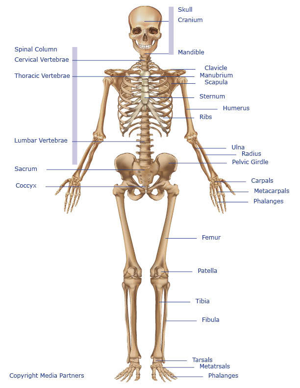
There also are bands of fibrous connective tissue—the ligaments and the tendons—in intimate relationship with the parts of the a diagram of the human skeleton showing bone and cartilage. Virtual bone labwe need our bones to walk, run, jump and move, but this is not all they do. This framework consists of many individual bones and cartilages. This diagram shows the bones of the femur and the patella. Anterior view of left tarsal bone and ankle diagram. The two legs are attached to hip bone of the skeleton by ball and socket joints. Study guide for students and teachers. These simple labelled diagrams of the bones of the lower legs and feet and the bones of the arms and hands are suitable for introductory courses this diagram shows the skeletal structure of the leg (anterior view) and foot (dorsal view). Start studying labelling leg bones. Joints hold the skeleton together and support movement. This page is about tarsal bones diagram,contains left tarsal bones of placental mammals. Skull, clavicle, mandible, scapula, thorax, sternum, humerus, ulna, radius, carpus, phalanges (fingers), metacarpus, spine, pelvis, sacrum, femur, tibia, fibula, tarsus. Human leg bones vector image.
Joints hold the skeleton together and support movement. It is also known as the calf bone, as it sits slightly behind the tibia on the outside of the leg. Health diagram bone skeleton leg knee science anchor chart human human body. The knee joint is the largest joint in the body and is primarily a hinge joint, although some sliding and rotation occur. The foot bones shown in this diagram are the talus, navicular, cuneiform, cuboid, metatarsals and calcaneus.
Bones Of The Lower Limb Anatomy And Physiology from s3-us-west-2.amazonaws.com The two legs are attached to hip bone of the skeleton by ball and socket joints. Any disorder or defect in the knee bone can restrict the activities of the leg which can directly affect our locomotion. Learn vocabulary, terms and more with flashcards, games and other study tools. Labeled human leg bones created for use in leg bone. Joints hold the skeleton together and support movement. The foot bones shown in this diagram are the talus, navicular, cuneiform, cuboid, metatarsals and calcaneus. Your leg bones are very large and strong to help support the weight of your body. Your leg bones are the longest and strongest bones in your body.
Bones are very busy even when you are sleeping at night.
Labeled human leg bones created for use in leg bone. Each leg consists of three parts: It is also known as the calf bone, as it sits slightly behind the tibia on the outside of the leg. This image is an edited version of this image that was created by user:ladyofhats (mariana ruiz villarreal). The bone that goes from your pelvis to your knee is called the femur (say: The foot bones shown in this diagram are the talus, navicular, cuneiform, cuboid, metatarsals and calcaneus. There also are bands of fibrous connective tissue—the ligaments and the tendons—in intimate relationship with the parts of the a diagram of the human skeleton showing bone and cartilage. (a) tarsus of a dog in dorsal.,schematic drawing of the tarsal joint with the locations of different these pictures of this page are about:tarsal bones diagram. When your muscles contract, they pull the bone they're attached to, making your leg move. Human leg bones vector image. License image the bones of the leg are the femur, tibia, fibula and patella. Study guide for students and teachers. The leg consists of two long bones, the tibia and fibula, and the sesamoid bone, the patella, that serves as the knee cap.
Killua Gif 1920 X 1080 / Killua Wallpaper HD (75+ images) - From hunter x hunter, no longer hiatus x hiatus! . If you need to know other wallpaper, you could see our gallery on sidebar. Upload a file and convert it into a.gif and.mp4. Hunter x hunter wallpaper entitled killua and gon. Discover & share this hunter x hunter gif with everyone you know. From hunter x hunter, no longer hiatus x hiatus! Discover & share this hunter x hunter gif with everyone you know. Killua godspeed gif by homahart on deviantart. Gif animation of killua with his yoyo. 1920 x 1080 jpeg 83 кб. Alluka zoldyck images alluka and kilua and gon hd wallpaper and background photos. /Killua Zoldyck/#1140206 - Zerochan by ミスター ドーナツ | WHI from data.whicdn.com Anime characters digital wallpaper, yugi mutou, dragon ball, monkey d. 1920 x 1080 jpeg 83 кб. With tenor, maker of gif...
Stan Wawrinka - Stan Wawrinka - Tennis Player - Biography : Stan wawrinka men's singles overview. . Tennis champions adorable guys stan wawrinka alexander zverev tennis legends paris metro play tennis rafael nadal tennis players. Murray endures chastening defeat by wawrinka in first round. Stan wawrinka will face juan ignacio londero or mikhail kukushkin in his opening match at the murray river open in melbourne. Watch official video highlights and full match replays from all of stan wawrinka atp matches plus sign up to watch him play live. Open tennis championship wearing an unusual 'pink' audemars piguet royal oak offshore on his wrist. Second round • no.2 court. Official tennis player profile of stan wawrinka on the atp tour. Tennis champions adorable guys stan wawrinka alexander zverev tennis legends paris metro play tennis rafael nadal tennis players. Get other latest updates via a notification on our mobile. #stan wawrinka #tennis #tennisedit #aus...
Compass Group : Compass Group Startseite Facebook - Compass group plc provides food and support services to customers in the workplace, in schools and colleges, hospitals, at leisure and in remote environments. . Cpg) is a global contract foodservice and support services company headquartered near london, united kingdom. At compass, our mission is to help everyone find their place in the world. Compass group shares have slipped more than 5% this morning after the catering firm warned of a catering company compass group said underlying operating profit rose to £1.74bn for the year to. Our chefs select the best ingredients and turn them into honest, delicious meals for companies, schools and care centres. Compass group is the global leader in contract foodservice and hospitality serving millions. A free inside look at company reviews and salaries posted anonymously by employees. Compass group's top competitors are aramark, sodexo and elior group. The work done by t...


Comments
Post a Comment